GYNEQUIPE
Monday to Thursday18:00 - 22:00
AddressXenias 1 & Vassilissis Sofias, Ilisia
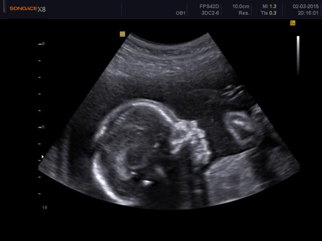
A second-trimester ultrasound, usually performed between the 18th and 23rd week of pregnancy, is a critical medical test that provides detailed information about the developing fetus and the mother’s reproductive system. This scan, also known as an ‘anatomy scan’ or ‘b-level ultrasound’, offers valuable information about the baby’s development, anatomy and overall health.
During the second trimester ultrasound, the gynecologist will place a gel on the mother’s abdomen and use a device (the ultrasound head) to send high-frequency sound waves through the uterus. These sound waves bounce off the baby and other structures, creating real-time images on a screen.
It should be noted here that these sound waves have no impact on the health of the baby and the woman.
In this way we consider:
Fetal anatomy: One of the primary goals is to assess the baby’s anatomy, including the brain, spine, heart, limbs, and organs. This helps detect any structural abnormalities or congenital problems.
Fetal measurements: The scan measures the baby’s size and growth, including head circumference, abdominal circumference and femur length, to determine whether growth corresponds to gestational age.
Amniotic fluid levels: The gynecologist checks the amniotic fluid levels around the baby, as too much or too little can indicate possible complications.
Locating the placenta: The position of the placenta is assessed to ensure that it is not covering the cervix (placenta previa), which can lead to complications during delivery.
The length of the cervix: The scan looks at the mother’s uterus and cervix for any abnormalities or potential problems that could affect the pregnancy.
Gender identification: If desired, the ultrasound can reveal the baby’s gender.
Confirmation of a multiple pregnancy: The scan can confirm whether the pregnancy includes one or more fetuses (twins, triplets, etc.).
Assessing blood flow: In some cases, the scan can assess blood flow in the umbilical cord and other major vessels to monitor the baby’s health.
We usually manage to see all the organs of the fetus with one examination, but this is not always possible, since the fetus in the mother’s abdomen constantly changes its position, with the result that some organs are not well visible. Otherwise, we repeat the examination later or on another day, completing the instruments that we did not see well the first time, due to position.
Just because an organ isn’t visible doesn’t mean it doesn’t exist.
The weight of the mother also plays an important role in this examination. In thin women, the ultrasound imaging is much more detailed and clear than in obese women. This can have implications for the diagnosis of various fetal problems.
In any case, however, future parents should know that under ideal examination conditions, the detection rate of anatomical anomalies does not exceed 70% of all organs examined, as very small malformations cannot be detected with modern ultrasound means. Also, some diseases, which are not accompanied by any anatomical abnormality and are called metabolic, cannot be detected by ultrasound.
In the event that an anatomical variation or other abnormality is detected, the gynecologist will advise the couple on the possibilities of prenatal testing, depending on the severity of the situation and they will decide together the treatment method.


Covering every aspect of a woman’s life

without stress and full of confidence during this special time

Minimally invasive surgery

One day diagnosis and treatment. (No touch, Diagnostic and Operative Hysteroscopy)

Personalized choice of treatment.

Urinary incontinence and pelvic prolapse treatment.

Prevention and treatment of gynaecological cancers. (International Guidelines)
Our services
Affiliated Hospitals
Affiliated Institutions
Office Hours
© Copyright 2024 Gynequipe. All right reserved

